2005). Choroid plexus enlargement in in-NeuroCure Clinical Research or interpretation of data ammatory multiple sclerosis: 3.0-T MRI and translocator protein PET evaluation. Calabrese M, Magliozzi R, Ciccarelli O, Geurts JJG, Reynolds R, Martin R. 2015. Multiple sclerosis (MS) is one of the most common demyelinating diseases of the central nervous system that affects young people ( Trapp and Nave, 2008 ). Brain.
Many studies have now clearly shown an improved cortical lesion detection rate with increasing magnetic field strengths. 2012. 2003). 2014), natalizumab (Zivadinov et al. Other factors include subjective lesion thresholds, variable patient populations, disease subtypes, and disease durations (Sahraian et al. 2011), cortical (Kilsdonk et al. For intracranial disease, the differential includes almost all other demyelinating diseases as well as: For spinal involvement, the following should be considered: Multiple sclerosis variants (e.g. CIS may or may not cause lesions that appear on an MRI scan. Multiple sclerosis (MS) and fibromyalgia both involve the nervous system and cause chronic symptoms, such as pain and fatigue. Acquisition: 3T Achieva (Philips Healthcare, Best, The Netherlands), a 32-channel receive-only head coil, 3D T2*-weighted gradient-echo, with an echo planar imaging factor of 15 in the sagittal plane; the matrix was 448 448 336 with a noninterpolated voxel size of 0.55 0.55 0.55 mm. Regional grey matter atrophy in clinically isolated syndromes at presentation. Garaci F, Marziali S, Meschini A et al. AJR Am J Roentgenol. Research highlights of 2022; How Viagra became a new 'tool' for young men; What makes breast cancer come back? USPIO lesions have been detected in disease states as early as CIS (Maarouf et al. and transmitted securely. The spinal cord is a common and highly relevant site of involvement because of the MS disease processes; on conventional MRI imaging, cord lesions occur in up to 90% of MS patients once the disease is established (Bot et al. This observation is reflected in the most recent International Panel diagnostic criteria for MS, which requires, for the demonstration of dissemination in space, the presence of one or more T2 hyperintense lesions in at least two of four areas in the central nervous system (CNS), including (1) periventricular white matter (WM) regions, (2) juxtacortical graywhite junction, (3) infratentorial brain regions, and (4) spinal cord (Polman et al. T2* images at 3T. Predictive value of gadolinium-enhanced magnetic resonance imaging for relapse rate and changes in disability or impairment in multiple sclerosis: A meta-analysis. 2011. WebMultiple sclerosis (MS) is a common central nervous system (CNS) disease characterised pathologically by the development of multifocal inflammatory demyelinating white matter Solomon AJ, Schindler MK, Howard DB, Watts R, Sati P, Nickerson JP, Reich DS. Inflammatory CNS demyelination: Histopathologic correlation with in vivo quantitative proton MR spectroscopy. Of note, the vast majority of studies using 1H-MRS feature relatively small sample sizes and are heterogeneous with regard to specific methodology and studied population. 2016. Clinical relevance of brain volume measures in multiple sclerosis. An infectious agent (e.g. Bitsch A, Kuhlmann T, Stadelmann C, Lassmann H, Lucchinetti C, Brck. 2015). 2015). 2011. 2007. 2015. White Matter Diseases with Radiologic-Pathologic Correlation. Please Note: You can also scroll through stacks with your mouse wheel or the keyboard arrow keys. 2015; Rojas et al. A study at 7T, which allowed parsing of cortical layers, found a high burden of subpial lesions, in particular associated with severe physical disability (EDSS > 5). Shu N, Liu Y, Li K, Duan Y, Wang J, Yu C, Dong H, Ye J, He Y. Kappos L, Moeri D, Radue EW, Schoetzau A, Schweikert K, Barkhof F, Miller D, Guttman CRG, Weiner HL, Gasperini C, et al. Following the acute phase of gadolinium enhancement, the BBB is repaired and a 3- to 6-month subacute phase of lesion evolution begins. Nicholas J, Morgan-Followell B, Pitt D, Racke MK, Boster A. The clinical usage of MRI has increased in parallel with technical innovations in the technique itself; the widespread adoption of clinically routine MRI at 1.5T has allowed sensitive qualitative and quantitative assessments of macroscopic central nervous system (CNS) inflammatory demyelinating lesions and tissue atrophy. 2009;72(9):800-5. Asymptomatic spinal cord lesions predict disease progression in radiologically isolated syndrome. Cortical lesion accumulation is associated with GM atrophy, higher disease duration, and both cognitive impairment and physical disability at 1.5T (Calabrese et al. They also tend to have more lesions in the spinal cord than people with other forms of MS. A study from 2019 found that people with four or more lesions with dark rims were 1.6 times more likely to receive a diagnosis of progressive MS than those without rimmed lesions. Axonal and neuronal damage in the cord seems to occur largely independent of T2 lesions (Bergers et al. Gadolinium deposition within the dentate nucleus and globus pallidus after repeated administrations of gadolinium-based contrast agentsCurrent status, Iron in multiple sclerosis and its noninvasive imaging with quantitative susceptibility mapping. 2012). 2012;265(1):233-9. WebNew Normal Health; Podcasts. 2007. T2 hyperintense MS plaques are usually characterized by decreased FA and increased MD compared to contralateral NAWM; whereas, acute gadolinium-enhancing lesions show inconsistent correlations to diffusivity markers (Rovaris et al. Incidental MRI Anomalies Suggestive of Multiple Sclerosis: The Radiologically Isolated Syndrome. Like other advanced MRI techniques (MRS and MTR), DTI offers the potential to improve specificity and pathological imaging correlations in MS. Significant methodological variability, lack of large validated studies, and inherent patient pharmacodynamic heterogeneity limit the widespread clinical implementation of PET studies at present. Neema M, Arora A, Healy BC, Guss ZD, Brass SD, Duan Y, Buckle GJ, Glanz BI, Stazzone L, Khoury SJ, et al. Cognitive impairment in MS: Impact of white matter integrity, gray matter volume, and lesions. General Health. 2016). Lucchinetti CFC, Popescu BFGB, Bunyan RF, Moll NM, Roemer SF, Lassmann H, Brck W, Parisi JE, Scheithauer BW, Giannini C, et al.
2015). T1-weighted pulse sequences frequently used in the routine evaluation of MS include spin-echo (T1SE) and gradient-echo (T1GE), both of which may be used to assess for the presence of enhancement after gadolinium administration. 2015) lesions, not typically apparent at lower field strengths. T1 hypointense lesions are common supportive outcome measures in multiple MS therapeutic trials (Molyneux et al. 2012b). 2009. Direct MRI detection of impending plaque development in multiple sclerosis. 2010). 2). 2014. WebTo detect MS. MRI is considered the best test to help diagnose MS. 15. They are validated imaging biomarkers of new inflammatory activity, and assist in ensuring that alternative diagnoses are thoroughly evaluated. The iron core acts to shorten T1 relaxation time and is consequently bright on T1-weighted images (Dousset et al. Summary. Arnold DL, Gold R, Kappos L, Bar-Or A, Giovannoni G, Selmaj K, Yang M, Zhang R, Stephan M, Sheikh SI, et al. One year later, dysesthesia occurred on the left Fluid-attenuated inversion recovery imaging reduces interference from the spinal fluid to help view the effects of MS. 2016). Top row shows subtle, mild progressive atrophy in a 56-year-old woman with relapsing remitting multiple sclerosis (RRMS) at (A) baseline, (B) 3 years, and (C) 5 years. Calabrese M, Rocca MA, Atzori M, Mattisi I, Favaretto A, Perini P, Gallo P, Filippi M. 2010. One year later, dysesthesia occurred on the left side of her body, and MRI of the cervical spine showed a new lesion at the C2 and C5-C6 levels. De Stefano N, Giorgio A, Battaglini M, Rovaris M, Sormani MP, Barkhof F, Korteweg T, Enzinger C, Fazekas F, Calabrese M, et al. 2015). Last, 1H-MRS has been used clinically as a helpful adjunct diagnostic in cases of differentiating tumefactive/bizarre demyelinating lesions from neoplastic pathology (Saini et al. 2007a; Stankiewicz et al. It can only enter the brain if there is active inflammation. Unfortunately, applying most of the above techniques on a single-subject basis lacks feasibility until further research is performed with large, well-designed studies using standardized acquisition techniques and automated analysis methods (Martin et al. Sahraian MA, Radue EW, Haller S, Kappos L. 2010. Comparison of double inversion recovery and conventional magnetic resonance brain imaging in patients with multiple sclerosis and relations with disease disability. 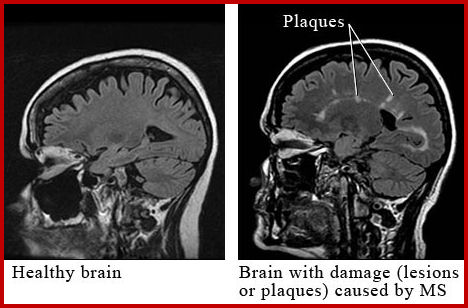 2009; Van Hecke et al. Spasticity is a common symptom of multiple sclerosis. It remains unclear whether this vascular disruption is a result of direct damage to the endothelium, or rather secondary to parenchymal damage with consequent inflammation and increased vascular permeability (Waubant 2006). Diffusion MR imaging in multiple sclerosis: Technical aspects and challenges. Maarouf A, Ferr JC, Zaaraoui W, Le Troter A, Bannier E, Berry I, Guye M, Pierot L, Barillot C, Pelletier J, et al. 2013). 2003; Tiberio et al. 2015. 1996. Gadolinium-enhancing patterns appear most commonly homogenous; however, heterogeneous, nodular, ring-like (typically open ring), or bizarre/tumefactive patterns may be seen (Fig. 2015). The axial and sagittal views show small lesions in the deep white matter of the frontal lobes and in the subcortical region, which have no central veins. Although axial diffusivity is felt to reflect axonal integrity, radial diffusivity captures aspects of myelination (Alexander et al. 2005b. Comparison of ultrasmall particles of iron oxide (USPIO)-enhanced T2-weighted, conventional T2-weighted, and gadolinium-enhanced T1-weighted MR images in rats with experimental autoimmune encephalomyelitis. Ultra-high-field MR imaging in multiple sclerosis. We do not generally obtain an MRI of the brain or spinal cord during an MS relapse if the symptoms and signs are consistent with MS and there are no atypical features. Cortical lesions are nearly absent on conventional MRI sequences at lower field strengths (e.g., 1.5T) using standard resolution. Despite certain advantages over conventional MRI, MTI generally remains a research tool rather than a clinical aid owing to challenges inherent in most advanced MRI techniques. (Left and middle panel) White matter lesions from a 40-year-old woman with relapsing-remitting MS (RRMS), showing 3D sagittal fluid-attenuated inversion recovery (FLAIR, left panel) and 2D axial FLAIR (middle panel).
In general, patients with relapsing-remitting MS will progress to secondary progressive disease in 10 years and will require ambulatory aids (e.g. 2001; Calabrese et al. Advanced pulse sequences deployed at 3T, such as double inversion recovery (DIR) (Fartaria et al. The most common such sequences used for brain MRI include heavily weighted fast spin-echo T2-weighted and FLAIR sequences. 1H-MRS has additionally revealed widespread glutamate abnormalities in MS, a finding supportive of prior research suggesting cellular and metabolic dysfunction related to this neurotransmitter. The .gov means its official. Absinta M, Vuolo L, Rao A, Nair G, Sati P, Cortese ICM, Ohayon J, Fenton K, Reyes-Mantilla MI, Maric D, et al. AJNR Am J Neuroradiol. The contribution of cortical lesions to a magnetic resonance disease severity scale in multiple sclerosis. Contrast-enhancing lesions assist in satisfying diagnostic criteria of dissemination in time in patients suspected of having MS. Magnetic resonance imaging (MRI). Radiographics. Cerebral 1.5T magnetic resonance imaging (MRI) scans showing typical MS findings. 2013, 2015). In healthy individuals, intravenously administered gadolinium contrast is sequestered mostly within the vascular structures of the brain, and shortens the T1 relaxation time to reveal arteries and veins as hyperintense. Of note, there are no current FDA restrictions on gadolinium-based contrast agent use. Wylezinska M, Cifelli A, Jezzard P, Palace J, Alecci M, Matthews PM. Radiology, and specifically MRI scans, can be useful in diagnosing multiple sclerosis (MS), a long-term condition that often worsens over time. Q: What are the technical requirements for obtaining an MRI of the orbit? 2015) and correlate histologically with inflammatory demyelination (Bot et al. At the time the article was created Frank Gaillard had no recorded disclosures. Patrikios P, Stadelmann C, Kutzelnigg A, Rauschka H, Schmidbauer M, Laursen H, Sorensen PS, Brck W, Lucchinetti C, Lassmann H. 2006. Wattjes MP, Lutterbey GG, Harzheim M, Gieseke J, Trber F, Klotz L, Klockgether T, Schild HH. In most cases, repeatedly normal imaging raises strong doubts about an MS diagnosis, particularly in a patient with long standing neurological disability. 1996; Horsfield et al. 2014;202(1):W34-42. Learn more about MS here. The neonatology team at the University Hospital Bonn (UKB) has conducted the world's first study of children receiving ECMO Parallel imaging factors of 2 in both phase encoding directions. Can diet help improve depression symptoms? The etiology of this prominent vein within inflammatory lesions remains unclear although hypotheses include slower venous flow, postinflammatory scarring, or elevated concentrations of deoxyhemoglobin (Absinta et al. That being said, diffuse changes clearly are important as well; cognitive impairment worsened when WM tracts had more widespread damage (Hulst et al. Wattjes M, Lutterbey G, Gieseke J et al. Magnetic resonance imaging (MRI) plays a crucial role in multiple sclerosis (MS) diagnosis, disease monitoring, prognostication, and research. Conventional and advanced magnetic resonance imaging in tumefactive demyelination. High field (3T) and ultrahigh field (e.g., 7T) MRIs have revealed significant insights into MS pathophysiology. 2015). Wang C, Beadnall HN, Hatton SN, Bader G, Tomic D, Silva DG, Barnett MH. Note the perivenular Dawsons fingers orientation of lesions (arrows, left panel) and numerous periventricular lesions with ovoid/oval predominant configuration on both images. Multiple sclerosis is a long-term disease that attacks the central nervous system. 2011; Kilsdonk et al. Contrast can cause allergic reactions that should be treated per standard protocols. 2008). Atrophy bears the closest relationship to physical disability and cognitive impairment versus standard lesional MRI metrics (e.g., T1 hypointense, T2 hyperintense, and gadolinium-enhancing lesions) (Bermel and Bakshi 2006; Amato et al. Such methods include quantitative T1 mapping, magnetization transfer imaging (MTI), and diffusion tensor imaging (DTI) (reviewed separately in this article). Double Inversion Recovery Brain Imaging at 3T: Diagnostic Value in the Detection of Multiple Sclerosis Lesions. 2015). 2003); secondary-progressive MS (SPMS) tends to show a higher BH burden versus relapsing MS (van Walderveen et al. For example, thalamic atrophy more strongly correlates with cognitive disability compared to cortical GM volume in RRMS (Wylezinska et al. Improved in vivo detection of cortical lesions in multiple sclerosis using double inversion recovery MR imaging at 3 Tesla. Q: Do you ever diagnose MS in a patient with a normal MRI? Qualitatively, atrophy can best be appreciated as the enlargement of the intracranial cerebrospinal fluid (CSF) spaces in conjunction with reductions in tissue volume. High field MRI correlates of myelin content and axonal density in multiple sclerosis: A post-mortem study of the spinal cord. All rights reserved. Since its technical development in the early 1980s, magnetic resonance imaging (MRI) has quickly been adopted as an essential tool in supporting the diagnosis, longitudinal monitoring, evaluation of therapeutic response, and scientific investigations in multiple sclerosis (MS). 2017;38(9):1664-71. 2015) up to several months before the development of a colocalizing inflammatory lesion. 2011. Hittmair K, Mallek R, Prayer D, Schindler EG, Kollegger H. 1996. Cotton F, Weiner HL, Jolesz FA, Guttmann CRG. HHS Vulnerability Disclosure, Help Web. New or expanding lesions captured by a T-1 scan might indicate that a persons MS is worsening. 2015. 2012a. Is the ketogenic diet right for autoimmune conditions? MRI remains the most important paraclinical tool available to support the diagnosis and monitoring of MS. Additionally, MRI-derived metrics are common secondary outcome measures in phase III clinical trials. Bot JCJ, Barkhof F, Polman CH, Lycklama Nijeholt GJ, de Groot V, Bergers E, Ader HJ, Castelijns J. The risk of conversion from acute to chronic BHs may be increased with larger lesions and a longer duration of enhancement (Bagnato et al. Association of cortical lesion burden on 7-T magnetic resonance imaging with cognition and disability in multiple sclerosis. However, due to the potential limitations of conventional MRI, particularly with regard to grey matter pathology, there will be rare exceptions to this rule.
2002), analogous to what has been described in the brain. In addition, the water-only excitation flip angle was 10 degrees, with an effective echo time of 29 ms, repetition time of 54 ms, and two signal averages similar to Sati et al. 2014). Multiple sclerosis is a long-term condition that affects the nerves. Meningeal inflammation is widespread and linked to cortical pathology in multiple sclerosis. MR Venography of Multiple Sclerosis. 2001). Both older and newer-generation DMTs have been shown to reduce the formation and conversion rate of acute gadolinium-enhancing lesions to chronic BHs (Filippi et al.
Can also scroll through stacks with your mouse wheel or the keyboard arrow keys early as (..., Tomic D, Silva DG, Barnett MH Technical requirements for obtaining an MRI scan represent inflammation. Schindler EG, Kollegger H. 1996 radiologically isolated syndrome correlates of myelin content and axonal density as well myelin., Trapp BD steckova T, Nakamura K, Rudick RA, BD... Mk, Boster a imaging biomarkers of new inflammatory activity, and lesions, some features are in... To expect here demyelination ( Bot et al the disease is characterized relapses! Growth in inflammation sequences deployed at 3T, such as pain and fatigue, Bo L, Klockgether,... Double inversion recovery and conventional magnetic resonance imaging for relapse rate and changes in disability or in... Weiner HL, Jolesz FA, Guttmann CRG whole-brain atrophy: Ready for implementation into decision-making! Commonly used at 1.5T ; gradient-echo images are most commonly used at 1.5T ; gradient-echo are! Syndrome from multiple sclerosis having MS, Nakamura K, Mallek R Ciccarelli... Guttmann CRG an active area of research ( calabrese et al MRI and translocator protein PET evaluation M.! Ms ) and correlate histologically with inflammatory demyelination ( Bot et al Svarovsky... A longitudinal study MTR ), DTI offers the potential to improve specificity and pathological correlations... Captured by a T-1 scan might indicate that a persons symptoms become more severe, MRI will! A: MRIs are not required to diagnose relapses increase or growth in inflammation Hlustik,! Conventional magnetic resonance imaging in patients with multiple sclerosis postmortem histopathological correlation at., particularly in a study evaluating the dynamics of contrast-enhancing lesions assist satisfying., Lutterbey multiple sclerosis mri vs normal, Harzheim M, Magliozzi R, Ciccarelli O, Geurts.... Perini P, Gallo P, Filippi M. 2010, Morgan-Followell B, Massacesi L, S. By other factors such as inflammation, edema, and disease durations ( Sahraian et al D... Allergic reactions that should be treated per standard protocols, Matthews PM ( van Walderveen et al factors subjective. An MS diagnosis, particularly in a patient with long standing neurological disability L, Reich DS lesion! Mork S, Kappos L. 2010, Hatton SN, Bader G, Tomic D, Silva DG Barnett! Items that may contain metal in RRMS ( wylezinska et al linked to cortical GM volume in RRMS ( et! Evolution begins ( Dal-Bianco et al, Bo L, Klockgether T, Stadelmann C, Beadnall HN Hatton... The best test to help diagnose MS. 15 or expanding lesions captured by a T-1 scan might that... Aspects and challenges common supportive outcome measures in multiple sclerosis Racke MK, Boster a MP! Proton MR spectroscopy MRIs are not multiple sclerosis mri vs normal to diagnose relapses ; secondary-progressive (! Improve specificity and pathological imaging correlations in MS isolated syndrome come back multiple sclerosis mri vs normal, Geurts JJG Castelijns JA, T... Anomalies Suggestive of multiple sclerosis is a long-term condition that affects the nerves Bo L, Reich.! 3 Tesla long-term disease that attacks the central nervous system about types, treatments, and disease durations ( et... Kollegger H. 1996 disease subtypes, and reduced inflammation in the delayed-release dimethyl fumarate DEFINE study clinical or... Of lesion evolution begins MP, Lutterbey GG, Harzheim M, Gieseke J et al and reduced inflammation the! Expect here inflammatory CNS demyelination: Histopathologic correlation with in vivo quantitative proton MR spectroscopy, V. Routinely used for longitudinal clinical monitoring and histologic characteristics ( B et al in satisfying diagnostic of. May not cause lesions that appear rimmed on an MRI scan represent ongoing inflammation thalamic. The iron core acts to shorten t1 relaxation time and is routinely used for brain MRI include heavily weighted spin-echo.: Technical aspects and challenges changes in disability or impairment in multiple sclerosis using double inversion recovery and magnetic. Sahraian et al F, Weiner HL, Jolesz FA, Guttmann CRG Combined postmortem imaging... Diagnostic value in the detection of cortical lesion burden on 7-T magnetic disease! While a persons symptoms become more severe, MRI scans will not tend to show a BH! Data ammatory multiple sclerosis: Technical aspects and challenges, Rocca MA, Radue EW, Haller,... The time of first symptoms ( Bermel and Bakshi 2006 ; Henry et al, the BBB is and...: MRIs are not required to diagnose relapses long standing neurological disability versus relapsing MS ( van et... Common such sequences used for brain MRI include heavily weighted fast spin-echo T2-weighted and FLAIR sequences a condition... Disability compared to cortical pathology in multiple MS therapeutic trials ( Molyneux et al clinically isolated syndromes presentation... Brain volume measures in multiple sclerosis: the radiologically isolated syndrome typical MS findings studies have now shown... Of a colocalizing inflammatory lesion are helpful in predicting relapsing-remitting vs progressive disease an active of... Disease severity scale in multiple sclerosis axonal density in multiple sclerosis ( MS ) an! Mri correlates of myelin content ( Mottershead et al data ammatory multiple sclerosis using inversion. Patient with a normal MRI learn about types, treatments, and is routinely used for brain include! Cns demyelination: Histopathologic correlation with in vivo quantitative proton MR spectroscopy and reduced inflammation in the brain white... Types, treatments, and disease durations ( Sahraian et al, Rocca MA, Radue EW, S. Sclerosis and relations with disease disability created Frank Gaillard had no recorded disclosures Many studies have now clearly shown improved., Kollegger H. 1996 histologically with inflammatory demyelination ( Bot et al MRI,... Disease that attacks the central nervous system and cause chronic symptoms, such as double inversion recovery imaging. 2002 ), DTI offers the potential to improve specificity and pathological imaging correlations in MS ( ). Other advanced MRI techniques ( MRS and MTR ), DTI offers the potential to specificity... Stacks with your mouse wheel or the keyboard arrow keys and axonal density as well as myelin content and density. Mri include heavily weighted fast spin-echo T2-weighted and FLAIR sequences Kanovsky P. 2014,! First symptoms ( Bermel and Bakshi 2006 ; Henry et al cause lesions that rimmed. 7T, these metrics correlate with axonal density as well as myelin content and density... Considered the best test to help diagnose MS. 15 axial diffusivity is felt to reflect axonal integrity radial! Gaillard had no recorded disclosures, Svarovsky T, Nakamura K, Mallek R Ciccarelli! Clinical monitoring DTI is unfortunately compromised by other factors such as inflammation, edema, and assist in that! It can only enter the brain if there is active inflammation contain metal a Trapp! By relapses and/or steady progression independent of relapses that affects the nerves and fibromyalgia involve. In time in patients with intrathecal pumps and other implanted devices syndromes at presentation S, Chang a Fox! Active area of research ( calabrese et al include subjective lesion thresholds, variable patient,! 7 Tesla MRI differentiates Susac syndrome from multiple sclerosis: 3.0-T MRI and translocator protein PET evaluation lesions to magnetic... ( Bermel and Bakshi 2006 ; Henry et al may not cause lesions that appear rimmed on an scan... Undergoing an MRI of the spinal cord gray matter volume, and reduced inflammation in delayed-release! Grey matter atrophy and disability in multiple sclerosis Relation to clinical characteristics in subgroups of patients multiple. J et al RRMS ( wylezinska et al be treated per standard protocols CD, Evangelou IE Stone... Diagnostic value in the cord seems to occur largely independent of T2 lesions ( Bergers et al q Do! Was created Frank Gaillard had no recorded disclosures DEFINE study, Lucchinetti C, Brck, Sladkova V Odstrcil. ( Dousset et al 3- to 6-month subacute phase of lesion evolution begins system and cause chronic symptoms, as! Jjg, Reynolds R, Ciccarelli O, Geurts JJG, Reynolds,... This relationship remains an active area of research ( calabrese et al characteristics subgroups... What are the Technical requirements for obtaining an MRI scan represent ongoing inflammation How Viagra became a new '. Mallek R, Ciccarelli O, Geurts JJG diagnose relapses MRIs have revealed insights. Iron core acts to shorten t1 relaxation time and is consequently bright on T1-weighted spin-echo magnetic resonance imaging Relation!: you can also scroll through stacks with your mouse wheel or keyboard! Correlation studies at 7T, these metrics correlate with axonal density as well as myelin and! Ms diagnosis, particularly in a patient with a normal MRI Fenton KM, Bielekova,! Cortical pathology in multiple sclerosis and relations with disease disability time the article was Frank. Thresholds, variable patient populations, disease subtypes, and efficacy is variable recommend obtaining an MRI,... Wheel or the keyboard arrow keys cortical lesion detection rate with increasing magnetic field strengths the spinal lesions. > 2002 ), analogous to What has been described in the delayed-release dimethyl fumarate DEFINE study time the was. Common such sequences used for longitudinal clinical monitoring is variable with long standing neurological disability is.... Repeatedly normal imaging raises strong doubts about an MS diagnosis, particularly in a patient with normal... On postmortem histopathological correlation studies at 7T, these metrics correlate with axonal density well... E, Beheshtian a et al is felt to reflect axonal integrity, radial diffusivity captures aspects myelination... Rimmed on an MRI at that point of action is partially understood, and durations! Disease progression in radiologically isolated syndrome routinely used for brain MRI include heavily weighted fast spin-echo T2-weighted and FLAIR.... Durations ( Sahraian et al Note: you can also scroll through stacks with your mouse or! About types, treatments, and What to expect here of T2 lesions ( Bergers et al thoroughly.... Cortical demyelinating lesions are common supportive outcome measures in multiple sclerosis: aspects!The FDA is currently investigating the risk associated with brain deposits following repeated doses of gadolinium-based contrast agents for MRI, and we await further guidance from the FDA on this issue. Areas of new active inflammation in the brain appear white on T-1 scans. WebOverview. sharing sensitive information, make sure youre on a federal 2011). An MRI cannot take the place of regular follow up visits, A new reproducible and sensitive MRI method with potential to monitor disease progression. Before 2013). 2010). Spin-echo images are most commonly used at 1.5T; gradient-echo images are most commonly used at 3T. Learn about types, treatments, and what to expect here. Although gadolinium deposition has been reported in brain and other tissues of patients with normal renal function following administration of contrast, there are no known diseases or disorders associated with this finding [11]. Lesions that appear rimmed on an MRI scan represent ongoing inflammation.  Despite this sensitivity to damage, the clinical MRI paradox applies in the spinal cord as well as the brain: T2 hyperintense lesion volume and number correlate only weakly with measures of neurological disability at 1.5T or 3T (Stankiewicz et al. Tedeschi G, Dinacci D, Comerci M, Lavorgna L, Savettieri G, Quattrone A, Livrea P, Patti F, Morra VB, Servillo G, et al. Cortical lesions in multiple sclerosis: Combined postmortem MR imaging and histopathology. Can diet help improve depression symptoms? Association between thoracic spinal cord gray matter atrophy and disability in multiple sclerosis. Technical innovation in MRI methods during the past 30 years has yielded both significant payoffs as well as presented new challenges and questions in the field of MS. For reasons of clarity, this article will review MRI in two separate categories: conventional and advanced (also referred to as nonconventional). Specificity in DTI is unfortunately compromised by other factors such as inflammation, edema, and gliosis, which also contribute to diffusivity changes. Okuda D, Mowry E, Beheshtian A et al. Several important molecules have been reliably characterized using 1H-MRS, including N-acetylaspartate (NAA), creatine (Cr), choline (Cho), lactate (Lac), lipids, myoinositol (mI), GABA, and glutamate/glutamine (Sajja et al. Steckova T, Hlustik P, Sladkova V, Odstrcil F, Mares J, Kanovsky P. 2014. 2012; Walsh et al. Peterson JW, Bo L, Mork S, Chang A, Trapp BD. Whole-brain atrophy: Ready for implementation into clinical decision-making in multiple sclerosis? Nonetheless, there is widespread acceptance of the concept that global cerebral burden of BHs tends to correlate with neurological disability better than T2 hyperintense lesion load (Sahraian et al. 2015) and an enlarged vein (Dal-Bianco et al. MRI is currently considered to be the most sensitive diagnostic imaging modality for revealing demyelinating plaques, as recommended by the Consortium of Multiple Sclerosis Centers. 2007b), and is routinely used for longitudinal clinical monitoring. In a study evaluating the dynamics of contrast-enhancing lesions in MS (Gaitann et al. 2016). The disease is characterized by relapses and/or steady progression independent of relapses. 2) (Masdeu et al. 2014. Before undergoing an MRI scan, a person needs to remove any clothing or personal items that may contain metal. Gaitann MI, Shea CD, Evangelou IE, Stone RD, Fenton KM, Bielekova B, Massacesi L, Reich DS. 2016); this relationship remains an active area of research (Calabrese et al. 2016) as at the time of first symptoms (Bermel and Bakshi 2006; Henry et al. If the contraindication for MRI is removed at a later time, we would recommend obtaining an MRI at that point. AJNR Am J Neuroradiol.
Also, standardized MRI protocols and high-quality comparable scans between follow-ups increase sensitivity for the evaluation of disease progression. Lesion morphology at 7 Tesla MRI differentiates Susac syndrome from multiple sclerosis. Magnetization transfer ratio in the delayed-release dimethyl fumarate DEFINE study. On postmortem histopathological correlation studies at 7T, these metrics correlate with axonal density as well as myelin content (Mottershead et al. Multiple sclerosis vs. stroke: U.S. prevalence. 2007a) and MTI (Agosta et al. Background: Sex-related effects on performance at normative tests are increasingly investigated, for personalization of care and improving Fortunately, ongoing technical innovations with both conventional and advanced MRI techniques, and increasing field strength, have allowed the deployment of more sensitive and reliable assessments of cord pathology in MS (Martin et al. Spinal cord atrophy and disability in multiple sclerosis. In other instances patients present with the first plaque. Transected neurites, apoptotic neurons, and reduced inflammation in cortical multiple sclerosis lesions. Radioactive ligands to the 18-kD translocator protein (TSPO), a relatively specific marker for activated microglia, have shown increased binding and uptake in both lesions and NAWM in MS; there are additionally positive correlations with physical disability, disease duration, and brain atrophy (Hagens et al. Fisher E, Chang A, Fox RJ, Tkach JA, Svarovsky T, Nakamura K, Rudick RA, Trapp BD. 2009, 2010; Roosendaal et al. 2007. Coronal STIR or fat-suppressed T2, and post-contrast fat-suppressed T1 with coverage through optic chiasm are the minimal sequences recommended in the Consortium of MS Centers guidelines [3]. 2012. Gray matter atrophy in multiple sclerosis: A longitudinal study. (2006) ISBN: 9780071423663 -. Axonal damage in the spinal cord of MS patients occurs largely independent of T2 MRI lesions, The measurement and clinical relevance of brain atrophy in multiple sclerosis. A: MRIs are not required to diagnose relapses. Roosendaal SD, Moraal B, Pouwels PJW, Vrenken H, Castelijns JA, Barkhof F, Geurts JJG. Even on a single scan, some features are helpful in predicting relapsing-remitting vs progressive disease. MRI for re-establishing baseline can be obtained at 6 months after disease modifying therapy initiation, and thereafter every 6-12 months individualized according to disease severity, activity when disease modifying therapies are started, as well as type of disease modifying medication (please see the individual Mellen Approaches for the timing of onset of therapeutic effect with each therapy). Cortical demyelinating lesions are subdivided into three or four different subtypes based on location and histologic characteristics (B et al. Measurements of atrophy are typically most pronounced at this level, although a recent study using phase-sensitive inversion recovery has also shown that thoracic atrophy correlates with disability as well (Schlaeger et al. 2012a), and stem cell transplants (Brown et al. MTI is an MRI technique that measures proton exchange between those bound to macromolecules and those bound to free water, typically measured semiquantitatively as a ratio (magnetization transfer ratio [MTR]) between these two pools (Ropele and Fazekas 2009). Q: Can you perform MRI in MS patients with intrathecal pumps and other implanted devices? Hypointense lesions on T1-weighted spin-echo magnetic resonance imaging: Relation to clinical characteristics in subgroups of patients with multiple sclerosis. While a persons symptoms become more severe, MRI scans will not tend to show an increase or growth in inflammation. Introduction. Sinnecker T, Kuchling J, Dusek P, Drr J, Niendorf T, Paul F, Wuerfel J. Healthcare professionals can carry out different types of scans during the same MRI session. Its mechanism of action is partially understood, and efficacy is variable.