The prognostic significance of sentinel node tumour burden in melanoma patients: an international, multicenter study of 1539 sentinel node-positive melanoma patients. Ann Surg Oncol 2018;25:210510. Epub 2022 Apr 19. Murali R, Desilva C, Thompson JF, Scolyer RA. Correspondence to The melanoma pathology report should include documentation of the features relied upon to establish a diagnosis of melanoma as well as features that are important for the prognosis and management of the patient.
For up to date recommendations, refer to Australian Cancer CouncilClinical practice guidelines for the diagnosis and management of melanoma. Mikael Hggstrm [note 1] Plast Reconstr Surg. breaking news vancouver, washington. Prognostic factors in cutaneous desmoplastic melanoma: a study of 252 patients. 2003;27:15716. Methods Mol Biol. Bruce R Smoller. Nevertheless, many additional well-established prognostic factors are not incorporated into the staging system. Arch Dermatol. It typically occurs in the head and neck region in severely sun-damaged skin of elderly patients. Melanoma in situ is often reported as a Clark level 1 melanoma. Web; . 2017;377:134556. It begins when the melanocytes in the skin grow out of control and form tumors. et al.
The presence of extranodal metastasis, although uncommon in SLNs, is also an adverse prognostic parameter; thus its presence or absence should be recorded in pathology reports of all regional lymph node specimens derived from melanoma cases [39]. Late regression is characterized by the presence of mature dermal fibrosis usually with accompanying loss of rete ridges in the overlying epidermis. Ann Surg Oncol. Adjuvant nivolumab versus ipilimumab in resected stage III or IV melanoma. The PubMed wordmark and PubMed logo are registered trademarks of the U.S. Department of Health and Human Services (HHS). T3, >2.04.0 mm. The two major forms of neurotropism are perineural invasion and intraneural invasion (Fig. 2014;106:djt435. WebWelcome to best cleaning company forever! You can see the main layers of the skin in this 2019;394(10197):471477. The goal of this chapter is to provide the reader with one perspective on the series of changes that are used in order to establish (or exclude) a diagnosis of melanoma. Punch scoring: a technique that facilitates melanoma diagnosis of clinically suspicious pigmented lesions. There were a number of reasons for removing mitotic rate as a staging parameter in the 8th edition. 3a). Griewank KG, Scolyer RA, Thompson JF, Flaherty KT, Schadendorf D, Murali R. Genetic alterations and personalized medicine in melanoma: progress and future prospects. Google Scholar. This article, attempts to describe the histologic features most closely associated with the various growth patterns of the most common subtypes of melanoma. The duration for which the lesion has been present and any history of recent change together with the clinical diagnosis or differential diagnosis may also be of assistance to the pathologist when interpreting the biopsy. Interobserver reproducibility of histopathologic prognostic variables in primary cutaneous melanomas. 2019;80:e1612. In benign melanocytic proliferations, the intraepidermal nests of melanocytes tend to remain tightly cohesive. The staging system is also important for eligibility, stratification, and analysis of clinical trials. Part I. 2017;377:181323. It is defined as a microscopic metastasis adjacent or deep to a primary tumor site identified on pathological examination. 2004;28:151825. In most nodular melanomas, however, the aggressive downward growth is apparent from the huge dermal nests and sheets of cytologically atypical melanocytes. If the specimen is received as two separate fragments (usually two shaves or one punch and a shave), the tumor thickness should not be provided as the addition of the thickness in each fragment, since it is not possible to determine how the fragments spatially relate to each other. In-situ lesions: 0.5 to 1 cm clinical margins Breslow depth less than 1 mm: 1 cm clinical margin Breslow depth between 1.01 to 2 mm: 1 to 2 cm clinical margin Am J Surg Pathol. J Am Acad Dermatol. In each of these cases, however, the Pagetoid cells are located in the central portions of the lesion and are relatively focal. Adjuvant dabrafenib plus trametinib in stage III BRAF-mutated melanoma. In the 8th edition, clinical staging is defined as being based upon assessment of the initial primary tumor biopsy as well as clinical examination of regional lymph nodes. Haydu LE, Holt PE, Karim RZ, Madronio CM, Thompson JF, Armstrong BK, et al. Pitfalls in Cutaneous Melanoma Diagnosis and the Need for New Reliable Markers, High regional mortality due to malignant melanoma in Eastern Finland may be explained by the increase in aggressive melanoma types, Current Trends of Immunotherapy in the Treatment of Cutaneous Melanoma: A Review, Reporting regression with melanoma in situ: reappraisal of a potential paradox, A retrospective study of malignant melanoma from a tertiary care centre in Saudi Arabia from 2004 to 2016, United States & Canadian Academy of Pathology Annual Meeting Abstracts. 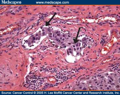
Nuclear chromatin is dense and nucleoli are often unapparent (Figure 8). T2, >1.02.0 mm. Tumor Size: This describes the size of the primary (original) tumor and whether it has invaded into nearby tissue. The previous minimum size and distance from the primary tumor that formed part of the 7th edition definition are not applicable in the 8th edition. High mitotic rate is an independent predictor of adverse outcome in melanoma patients. doi: 10.1097/00000658-199309000-00005. 2). As is commonly observed clinically in primary melanomas, the immune system can react against a primary melanoma and result in loss of part or all of the tumor. Ingrid Ferreira, Alastair Droop, David J. Adams, Emily L. Clarke, Ryckie G. Wade, Darren Treanor, Richard A. Scolyer, Robert V. Rawson, Victor G. Prieto, Magdalena Ciyska, Grayna Kamiska-Winciorek, Aleksandra Lesiak, Modern Pathology The mucosal surface is often spongiotic and may be acanthotic. doi: 10.1001/archsurg.1991.01410280036004. The T category is divided into T1T4 based on the tumor thickness. Melanoma in situ Tis is used to designate melanoma in situ. Characteristics, treatment and outcomes of 589 melanoma patients documented by 27 general practitioners on the Skin Cancer Audit Research Database. The various N categories are presented in Table3. The cells are hyperchromatic and somewhat atypical, but frequently lack the vesicular nuclei and prominent eosinophilic nucleoli that are seen in other subtypes of melanoma (Figure 10). More accurate personalized predication of prognosis is likely to be possible in the future utilizing web-based or other computerized tools, the integration of additional prognostic factors and complex molecular data as well as molecular predictive and diagnostic biomarkers. In superficial spreading melanomas, abundant single melanocytes are present within the epidermis (Figure 1). Mitotic rate was removed as a T1 subcategory criterion in the 8th edition. Lentigo maligna melanoma is, by definition, a melanoma that invades the dermis. 2019 Jul;81(1):204-212. doi: 10.1016/j.jaad.2019.01.051. a LM with, Histologic appearance of LM compared to non-LM melanoma in situ. The New Zealand Cancer Registry does not publish the figures for melanoma in situ, but unpublished data suggest that about the same number of people are diagnosed with in-situ melanoma as those diagnosed with invasive melanoma [2]. Primary melanoma of the skin: recognition and management. Available at: Higgins HW 2nd, Lee KC, Galan A, Leffell DJ. The dermal component of acral lentiginous melanoma generally demonstrates fascicles of spindle-shaped melanocytes that may course within fibrotic stroma. <2 or 3 mm but not continuous with edge: "Close margins at __ mm at (location). In general terms, melanoma in situ is macular (flat). The most common subtypes are: Rare forms of melanoma that may have an in-situ phase include: There were 2423 melanoma registrations in New Zealand in 2015. [note 5], For a full list of contributors, see article. Lancet. There is a comprehensive literature that critically evaluates histologic parameters associated with this collection of tumors and relates them to prognostic information, and no attempt will be made to correlate the histologic change with prognostic information. National Library of Medicine Melanoma Institute Australia, The University of Sydney, Sydney, NSW, Australia, Richard A. Scolyer,Robert V. Rawson&Peter M. Ferguson, Sydney Medical School, The University of Sydney, Sydney, NSW, Australia, Tissue Pathology and Diagnostic Oncology, Royal Prince Alfred Hospital & NSW Health Pathology, Camperdown, NSW, 2050, Australia, University of Texas, MD Anderson Cancer Center, Houston, TX, 77030, USA, Jeffrey E. Gershenwald&Victor G. Prieto, You can also search for this author in Diagnostic histological criteria are best organized by architectural and cytologic features, and examined in terms of epidermal findings and dermal findings. Scolyer, R.A., Rawson, R.V., Gershenwald, J.E. Dodds TJ, Lo S, Jackett L, Nieweg O, Thompson JF, Scolyer RA. Underpinned by improved understanding of the molecular basis of melanoma and regulation of immune system [7], new effective targeted and immune therapies have transformed the management of patients with widespread melanoma metastases. In November 2015, the International Melanoma Pathology Study Group (IMPSG) met at the University of California, San Francisco, and considered, discussed, debated, and voted upon various pathology staging issues. Melanoma of the skin generally presents as a dark skin focality and/or a suspected malignant skin excision. A brisk host response is present underlying a small focus of dermal invasion in this superficial spreading type of melanoma. Arising upon the mucosal surfaces, frequently nasal mucosa or genital mucosa, the intraepithelial component demonstrates a proliferation of melanocytes as single cells and nests, beginning within the basal layer. 3b). In concert with individual melanocytes becoming smaller with progressive descent, the nesting pattern of these cells also changes reproducibly within benign nevi. It is also known as in-situ melanoma and level 1 melanoma. Clinical appearance of LM compared to non-LM melanoma in situ. Another relatively common subtype of melanoma is the nodular melanoma. Eye (Lond). Similarly, a melanoma measuring 1.04mm thick would be recorded as 1.0mm in the pathology report and designated as T1b for staging. Yes, left untreated, in situ can grow and reach the vascular level where it can morph into something else and has a method of transport to distant areas.but in and of itself at first recognition,in situ is NOT melanoma.